Somatotopic map
Somatotopic map of the preterm sensorimotor cortex
In the mature brain, the primary somatosensory and motor cortices are known spatially organized such that neural activity relating to specific body parts can be somatopically mapped onto an anatomical “homunculus”. We characterized the somatotopic organization of the primary sensorimotor cortices in 35 healthy preterm infants aged from 31+6 to 36+3 weeks postmenstrual age using functional MRI and a set of custom-made robotic tools (to stimulate the wrists, ankles and mouth).
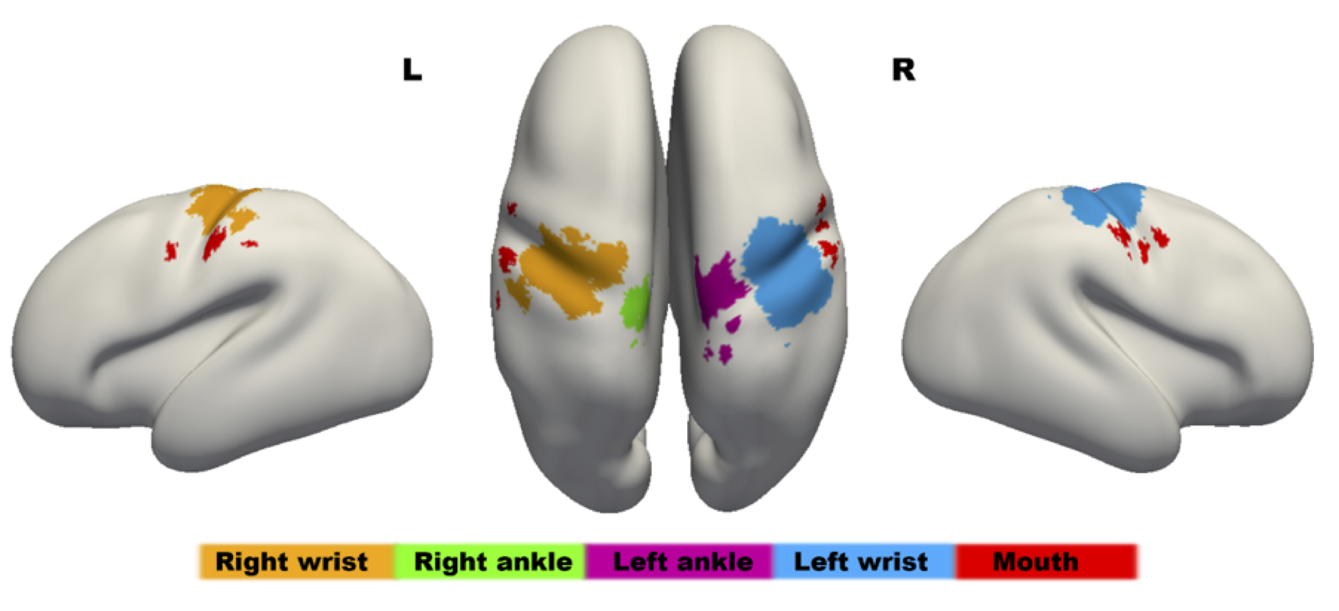
Figure: The sensorimotor homunculus in the preterm human brain at 34 weeks PMA. The map has been overlaid onto an age-specific inflated brain template using a “winner-takes-all” approach after combing the significant results of the group level activation maps from each stimulated body part. In agreement with the well characterized adult somatotopic map, functional activity relating to the ankles (green and purple) is located superiorly to those of the wrist (orange and blue) and mouth (red).
Download
The activation maps represent the results of one sample t-tests using permutation testing and threshold free cluster enhancement (image values are 1-p format (ie: to threshold at p<0.05, minimum signal intensity should be set to 0.95)). The maps have been aligned to the 34 week PMA templates available for download on this site. Please download the data from here.
For individual subject data please contact tomoki.arichi@kcl.ac.uk
Material
The characteristics of the infants studied to produce this activation maps are described in the following table:
| Body part group | n | GA at birth in weeks median (range) | Birth weight in grams
median (range) |
PMA at scan in weeks median (range) |
| Left ankle | 10 | 34+2 (28+3 – 36+1) | 1850 (1120 – 3110) | 35+2 (33+6 – 36+3) |
| Left wrist | 10 | 32+3 (26+3 – 34+5) | 1930 (840 – 2330) | 34+2 (33+0 – 35+3) |
| Right ankle | 9 | 33+3 (28+5 – 35+4) | 2100 (1340– 3110) | 34+4 (31+6 – 36+3) |
| Right wrist | 10 | 32+4 (29+1 – 35+6) | 1680 (1330-2100) | 34+2 (33+3 – 36+1) |
| Mouth | 10 | 34+1 (30+4 – 35+4) | 1980 (1440 – 3110) | 35+1 (32+3 – 36+3) |
Methods
MR images were collected following feeding and during natural sleep on a 3-Tesla MRI scanner. Blood Oxygen Level Dependent (BOLD) functional MRI data (256 volumes) were collected using a T2*-weighted single-shot gradient echo echo-planar imaging (GRE-EPI) sequence (resolution: 2.5*2.5*3.25mm; 21 slices; TE: 45msec; TR: 1500msec, FA: 90 degrees; SENSE factor 2). Somatosensory stimulation was provided by a set of automated MR compatible robotic tools which induced 0.33Hz flexion/extension movement of the wrists and ankles for 24 seconds interleaved with 24 seconds of rest. Stimulation of the mouth was achieved using a puffs of air (0.4atm at 0.3Hz) on the lips and mouth for 24 seconds interleaved with 24 seconds of rest.
The study and methods are described in detail in the following publication:
Sofia Dall’Orso, Johannes K Steinweg, Alessandro G Allievi, A David Edwards, Etienne Burdet, Tomoki Arichi. Somatotopic mapping of the developing sensorimotor cortex in the preterm human brain. Cerebral Cortex 2018 (in press)